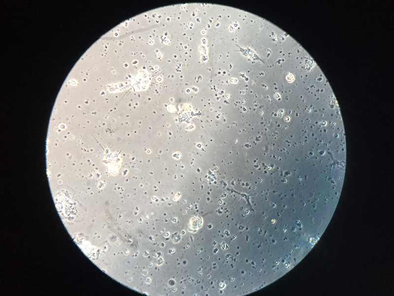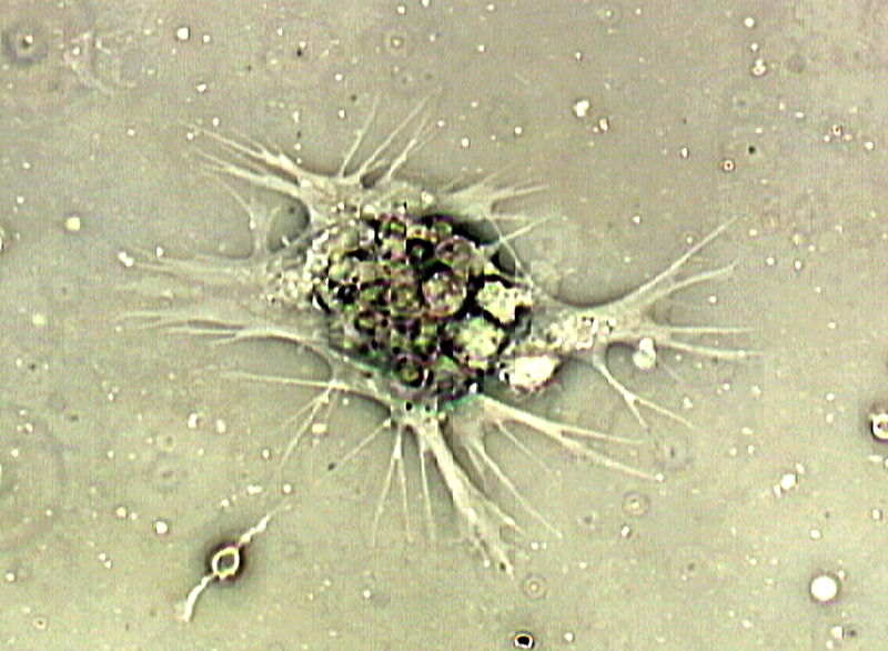
Hunting for Hemocytes Laboratory
Form, function, and microscopy
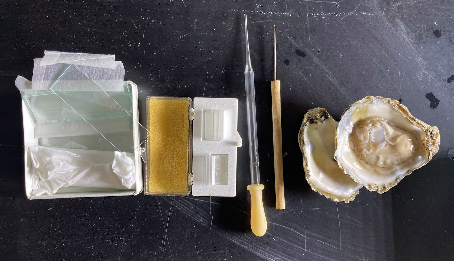
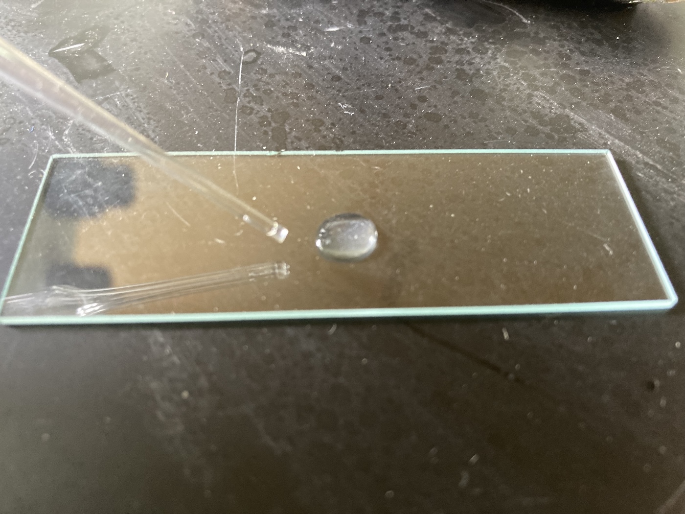
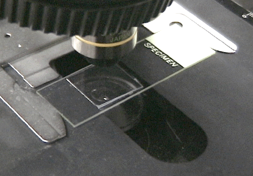
Materials
This activity uses:
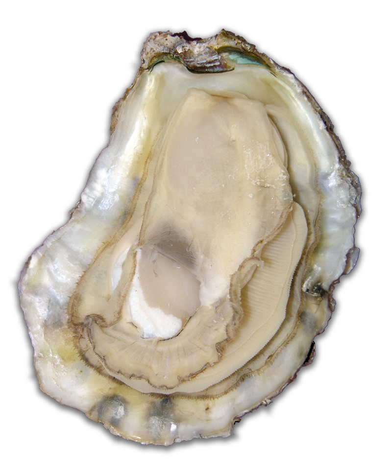
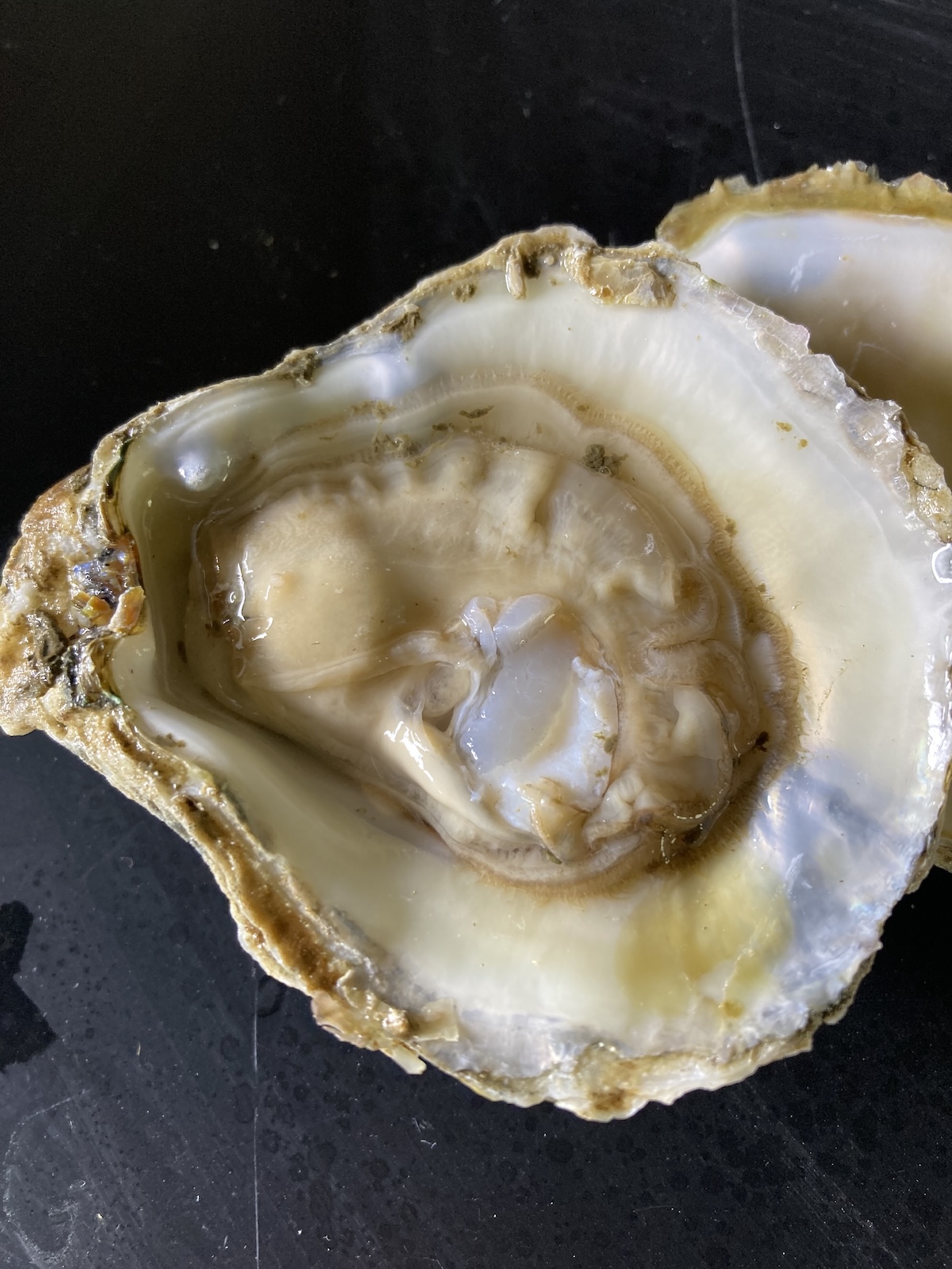
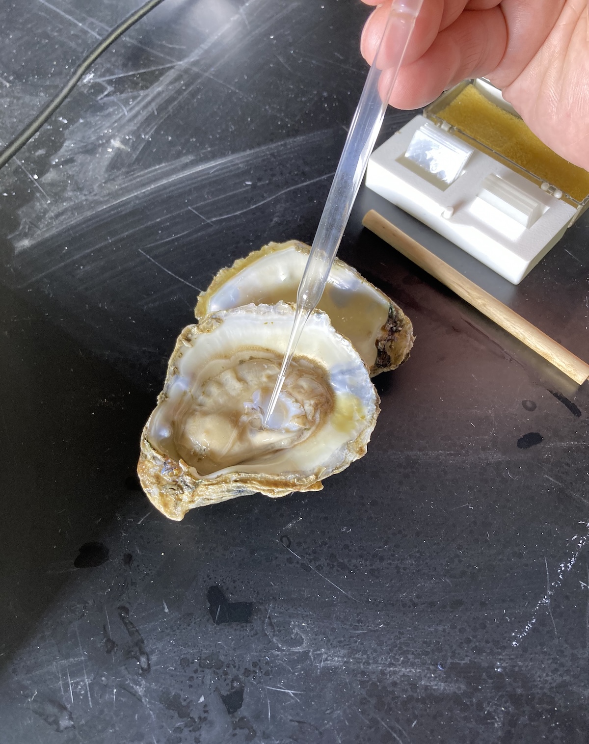
- Live oysters
- Shallow glass or plastic dishes
- Compound light microscope with 10X, 20X and 40X objectives
- Glass slides and glass cover slips
- Probe
- Glass capillary pipette, micropipette or glass eyedropper