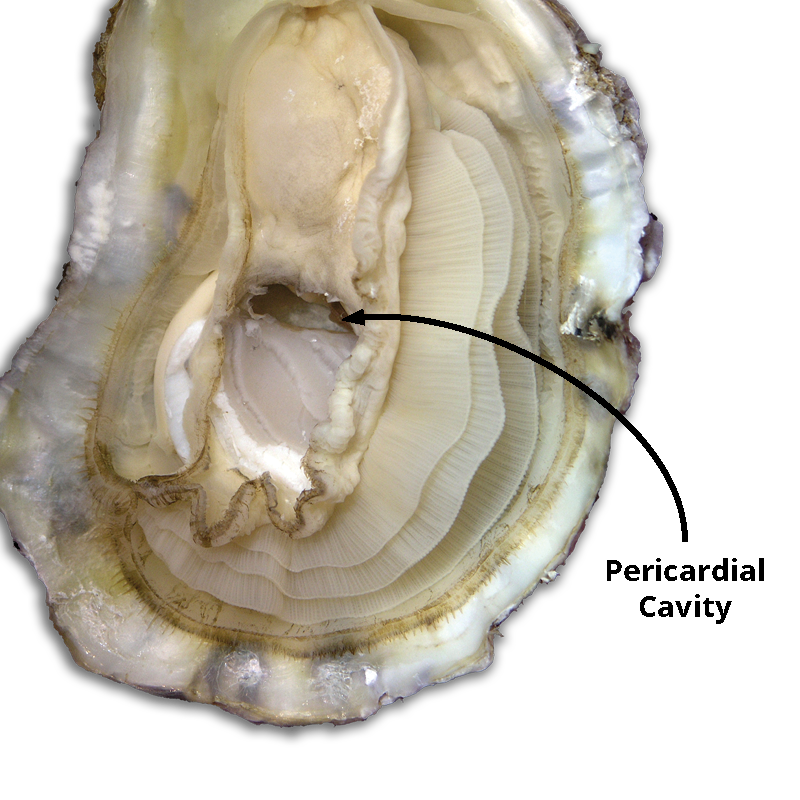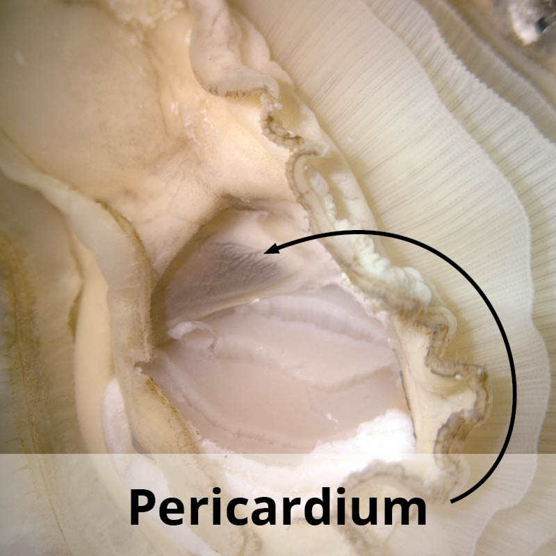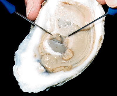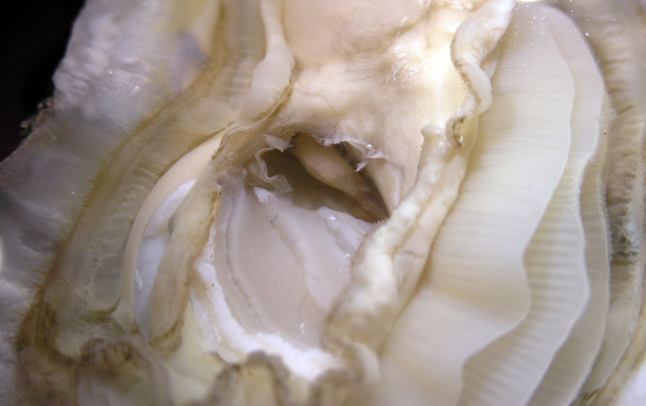
Oyster Anatomy Laboratory
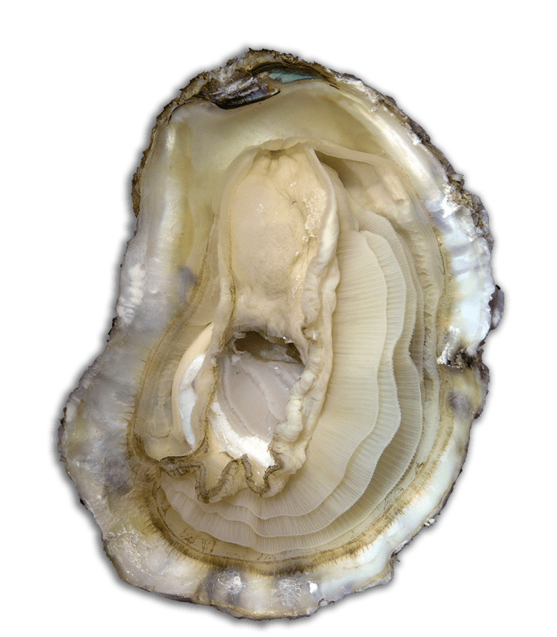
Internal Anatomy: Observation and Investigation
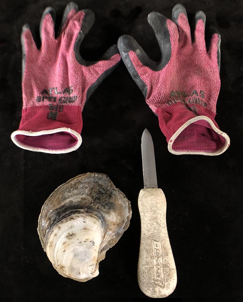
Materials
For Lab:
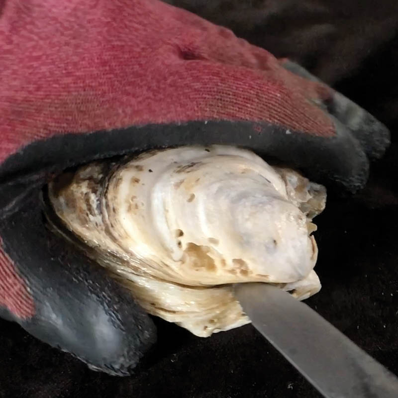
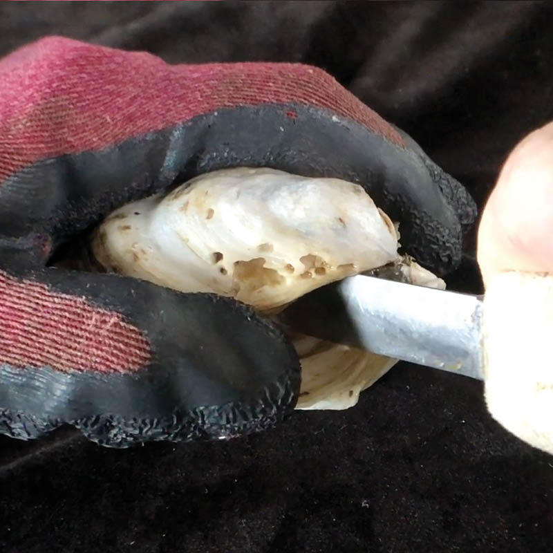
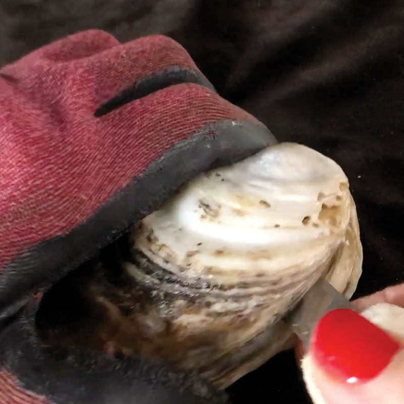
- Oyster knife
- Sturdy gloves
- Dissecting kit
- Artificial saltwater (10-15ppt)
- Computer access
Per Lab Group:
- Live oysters
- Shallow glass or plastic dishes (min 2” depth)
- Probes
- Forceps
- Computer access
- Internal Anatomy Handout (1 per student)
- Stereomicroscopes (optional)
- Large magnifying glasses (optional)